The Biological Context: Why Bacteria “Stick”
To understand urinary tract health, we must first understand the persistence of the organisms that challenge it. By far the most common culprit in canine urinary issues is Escherichia coli (E. coli).
Under a microscope, these bacteria are not merely floating passive cells. They are equipped with specialized, hair-like appendages called Type 1 Fimbriae. Think of these fimbriae as microscopic grappling hooks or Velcro.
When bacteria enter the bladder, the body’s natural defense is urination — a powerful mechanical flush designed to wash intruders out. However, the Type 1 fimbriae allow the bacteria to latch onto the mannosylated receptors on the epithelial lining of the bladder wall. Once anchored, they are resistant to the flow of urine, allowing them to colonize and create the irritation characteristic of urinary tract challenges.
The Science of D-Mannose: Competitive Inhibition
D-Mannose is a simple sugar isomer chemically related to glucose, but it functions very differently in the body. It does not behave as a fuel source; instead, it acts as a molecular decoy.
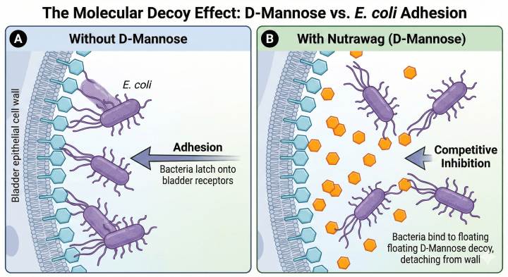
The mechanism at work is known as Competitive Inhibition.
The receptors on the bladder wall that bacteria target are composed of D-Mannose molecules. When you introduce free-floating D-Mannose into the urine (via supplementation), you flood the bladder with these “decoy” targets.
Because the bacteria have a high affinity for D-Mannose, their fimbriae bind to the free-floating D-Mannose molecules suspended in the urine rather than the D-Mannose structures attached to the bladder wall. Essentially, the bacteria “let go” of the tissue to grab the supplement. Once bound to the free D-Mannose, the bacteria can no longer anchor themselves to the lining.
What the Clinical Data Says
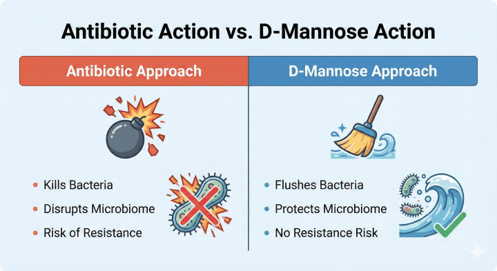
Clinical research distinguishes D-Mannose from traditional antimicrobial agents because its action is mechanical, not bactericidal.
- No “Die-Off” Stress: Unlike antibiotics, which rupture bacterial cell walls, D-Mannose does not kill the bacteria. It simply inhibits their adhesion.
- Resistance Neutral: Because D-Mannose does not attack the bacteria’s survival mechanisms, the bacteria are under no evolutionary pressure to mutate or develop resistance. This makes it a sustainable option for long-term maintenance.
- Preservation of Microbiome: Studies suggest that because D-Mannose is specific to the Type 1 fimbriae of E. coli and similar organisms, it does not indiscriminately wipe out beneficial gut or bladder flora, unlike broad-spectrum interventions.
Why NutraWag Chose This Ingredient
We selected D-Mannose as the “Anchor Ingredient” for our urinary formula because it addresses the root cause of persistence — adhesion — without the collateral damage associated with harsh antimicrobials.
Many supplements rely on low-dose cranberry powder alone, which acts via a similar anti-adhesion mechanism (PACs) but often lacks the concentration required to saturate the urine. By utilizing D-Mannose as a primary agent, NutraWag ensures there is a sufficient concentration of “decoys” in the urine to support the body’s natural ability to flush impurities during regular voiding.
Clinical Summary
- Adhesion is the Enemy: Bacteria survive in the bladder by using “grappling hooks” (Type 1 fimbriae) to cling to the wall.
- The Decoy Effect: D-Mannose acts as a molecular decoy, tricking bacteria into binding to the urine instead of the bladder lining.
- Mechanical Flushing: Once bound to D-Mannose, bacteria are rinsed away naturally during urination.
- Microbiome Safe: Because it doesn’t kill bacteria, D-Mannose supports urinary health without disrupting the dog’s healthy gut flora.
References
Scientific References
- Altarac, S., & Papeš, D. (2014). Use of D-mannose in prophylaxis of recurrent urinary tract infections (UTIs) in women. BJU International, 113(1), 9–10.
Note: This is a key human clinical study often cited regarding D-mannose’s ability to reduce the risk of recurrent UTIs by inhibiting bacterial adherence. - Domenici, L., Monti, M., Bracchi, C., Giorgini, M., Colagiovanni, V., Muzii, L., & Benedetti Panici, P. (2016). D-mannose: a promising support for acute urinary tract infections in women. A pilot study. European Review for Medical and Pharmacological Sciences, 20(13), 2920–2925.
Note: This pilot study supports D-mannose as a safe and effective management tool for acute UTIs, highlighting its mechanism of action. - Mulvey, M. A., Lopez-Boado, Y. S., Wilson, C. L., Roth, R., Parks, W. C., Heuser, J., & Hultgren, S. J. (1998). Induction and evasion of host defenses by type 1-piliated uropathogenic Escherichia coli. Science, 282(5393), 1494–1497.
Note: A seminal foundational paper explaining the biology of Type 1 fimbriae and how bacteria use them to attach to bladder cells. - Scribano, F., Marchetti, P., & Mura, U. (2021). Considerations on D-mannose Mechanism of Action and Consequent Classification of Marketed Healthcare Products. Frontiers in Pharmacology, 12, 636377.
Note: An excellent review article that details the non-pharmacological, mechanical “competitive inhibition” action of D-mannose. - Sokurenko, E. V., Vogel, V., & Thomas, W. E. (2008). Catch-bond mechanism of force-enhanced adhesion by bacteria. Cell, 135(2), 237–246.
Note: A deep scientific dive into the biophysics of how bacterial FimH adhesins (the “hooks”) bind to mannose receptors.
Disclaimer: While many fundamental mechanisms of action are conserved across species, some citations refer to human clinical studies or in vitro (test tube) models. These references are provided for educational purposes regarding the underlying biological mechanisms of ingredients and are not intended to imply specific clinical outcomes in dogs.


